MedGemma 1.5 Technical Report
arXiv:2604.05081v2 · This workspace stays on this version. Check arXiv for newer versions
Some source details need review 3 to check
Original formulas, table values and figures are preserved. Comparison can miss differences; use the original PDF to check details.
Reading view checked: 18 math expressions, 1 rendering failures; 16 of 16 images available.
Compared 123 of 123 source passages against 23 of 23 PDF pages.
- Some formulas could not be rendered; their original TeX is shown. Check the original PDF. (1)
HTML S2_T2 says “MS-CXR-T (Bannur et al., 2023b),” while PDF page 5 says “MS-CXR-T (Bannur et al., 2023a),” so the PDF contradicts the HTML. Check PDF page 5
PDF page 9 reads “(Bannur et al., 2023a),” while HTML block S3_SS2_SSS3_p1_1 reads “([Bannur et al., 2023b](#bib.bib22)).” Check PDF page 9
16 of 16 figure, table and graphic images captured. Open original PDF
Abstract
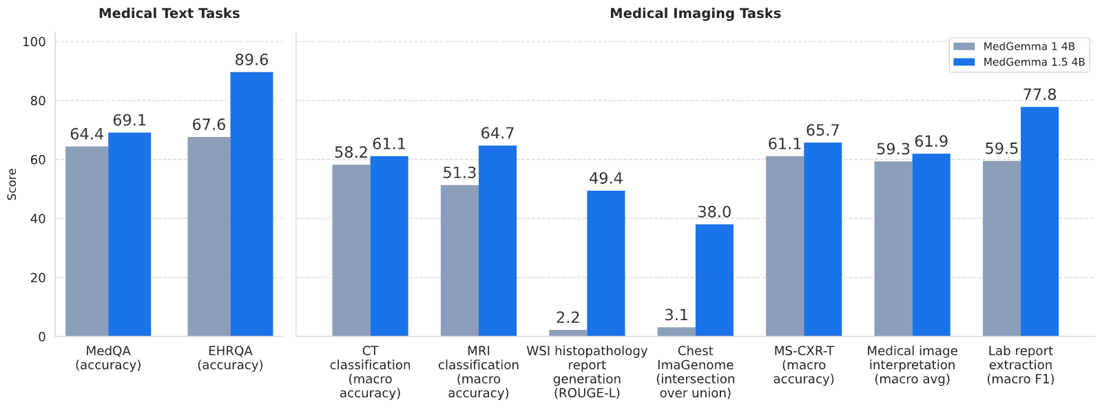
We introduce MedGemma 1.5 4B, the latest model in the MedGemma collection. MedGemma 1.5 expands on MedGemma 1 by integrating additional capabilities: high-dimensional medical imaging (CT/MRI volumes and histopathology whole slide images), anatomical localization via bounding boxes, multi-timepoint chest X-ray analysis, and improved medical document understanding (lab reports, electronic health records). We detail the innovations required to enable these modalities within a single architecture, including new training data, long-context 3D volume slicing, and whole-slide pathology sampling. Compared to MedGemma 1 4B, MedGemma 1.5 4B demonstrates significant gains in these new areas, improving 3D MRI condition classification accuracy by 11% and 3D CT condition classification by 3% (absolute improvements). In whole slide pathology imaging, MedGemma 1.5 4B achieves a 47% macro F1 gain. Additionally, it improves anatomical localization with a 35% increase in Intersection over Union on chest X-rays and achieves a 4% macro accuracy for longitudinal (multi-timepoint) chest x-ray analysis. Beyond its improved multimodal performance over MedGemma 1, MedGemma 1.5 improves on text-based clinical knowledge and reasoning, improving by 5% on MedQA accuracy and 22% on EHRQA accuracy. It also achieves an average of 18% macro F1 on 4 different lab report information extraction datasets (EHR Datasets 2, 3, 4, and Mendeley Clinical Laboratory Test Reports). Taken together, MedGemma 1.5 serves as a robust, open resource for the community, designed as an improved foundation on which developers can create the next generation of medical AI systems. Resources and tutorials for building upon MedGemma 1.5 can be found at https://goo.gle/medgemma.
Expanding the capabilities of open-weight medical foundation models to encompass complex, high-dimensional modalities is essential for comprehensive healthcare AI development. While recent advancements (Yang et al., 2024; Sellergren et al., 2025) have demonstrated the utility of multimodal models in standard 2D imaging tasks, the number of models and benchmarks addressing more complicated imaging tasks are more limited. In this technical report, we introduce MedGemma 1.5, an updated model in the MedGemma collection integrating support for high-dimensional, volumetric, and longitudinal data, along with existing support for 2D imaging and text-based knowledge and reasoning, all within a single unified architecture.
Specifically, MedGemma 1.5 expands the multimodal capabilities of previous releases by introducing native support for four medical imaging capabilities including: (1) 3D radiology interpretation (both CT and MRI volumes), (2) whole slide image (WSI) interepretaion for histopathology, (3) fine-grained anatomical localization for X-rays via bounding boxes, and (4) multi-timepoint radiology analysis. Through additional curated training datasets, we also incorporated improved capabilities for medical document (PDF) understanding and improved text-based clinical reasoning. As the first open model to achieve these diverse baseline capabilities in a single architecture, MedGemma 1.5 serves as a robust, open resource for the community, designed as an improved foundation on which developers can create the next generation of medical AI systems.
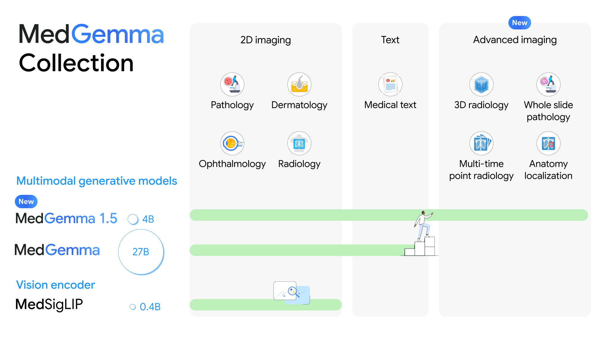
Similar to MedGemma 1, MedGemma 4B 1.5 is based off of Gemma3 (Team et al., 2025) with the same architecture. The vision encoder is 400M MedSigLIP encoder (Sellergren et al., 2025).
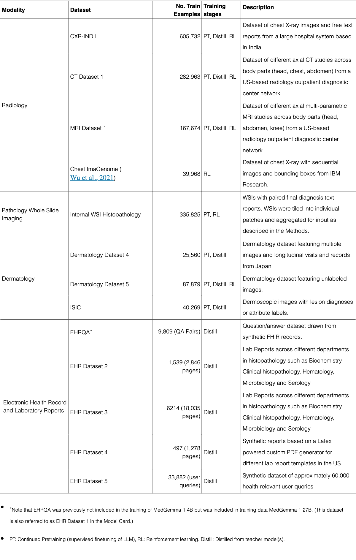
The methodological updates for MedGemma 4B version 1.5 primarily fall into three categories: first, we incorporated several new training datasets with the goal of broadening medical knowledge and expanding capabilities for “high-dimensional” medical imaging including 3D CT and MRI volumes and Whole Slide Images (WSIs) for histopathology. Second, we implemented minor modifications to the modeling methodology to improve training efficiency and model capabilities. Third, we expanded evaluations to assess model performance on updated capabilities.
| Modality | Dataset | No. Train Examples | Training stages | Description |
| Radiology | CXR-IND1 | 605,732 | PT, Distill, RL | Dataset of chest X-ray images and free text reports from a large hospital system based in India |
| CT Dataset 1 | 282,963 | PT, Distill, RL | Dataset of different axial CT studies across body parts (head, chest, abdomen) from a US-based radiology outpatient diagnostic center network. | |
| MRI Dataset 1 | 167,674 | PT, Distill, RL | Dataset of different axial multi-parametric MRI studies across body parts (head, abdomen, knee) from a US-based radiology outpatient diagnostic center network. | |
| Chest ImaGenome (Wu et al., 2021) | 39,968 | RL | Dataset of chest X-ray with sequential images and bounding boxes from IBM Research. | |
| Pathology Whole Slide Imaging | Internal WSI Histopathology | 335,825 | PT, RL | WSIs with paired final diagnosis text reports. WSIs were tiled into individual patches and aggregated for input as described in the Methods. |
| Dermatology | Dermatology Dataset 4 | 25,560 | PT, Distill | Dermatology dataset featuring multiple images and longitudinal visits and records from Japan. |
| Dermatology Dataset 5 | 87,879 | PT, Distill, RL | Dermatology dataset featuring unlabeled images. | |
| ISIC | 40,269 | PT, Distill | Dermoscopic images with lesion diagnoses or attribute labels. | |
| Electronic Health Record and Laboratory Reports | EHRQA∗ | 9,809 (QA Pairs) | Distill | Question/answer dataset drawn from synthetic FHIR records. |
| EHR Dataset 2 | 1,539 (2,846 pages) | Distill | Lab Reports across different departments in histopathology such as Biochemistry, Clinical histopathology, Hematology, Microbiology and Serology | |
| EHR Dataset 3 | 6214 (18,035 pages) | Distill | Lab Reports across different departments in histopathology such as Biochemistry, Clinical histopathology, Hematology, Microbiology and Serology | |
| EHR Dataset 4 | 497 (1,278 pages) | Distill | Synthetic reports based on a Latex powered custom PDF generator for different lab report templates in the US | |
| EHR Dataset 5 | 33,882 (user queries) | Distill | Synthetic dataset of approximately 60,000 health-relevant user queries |
View original table
| Task | Dataset | Modality | No. Eval. Examples | OOD | Public |
| Medical text QA | EHRNoteQA (Kweon et al., 2024) | Text | 962 | ✓ | ✓ |
| CXR temporal analysis | MS-CXR-T (Bannur et al., 2023b) | Radiology longitudinal | 1326 | - | ✓ |
| 3D CT classification | CT Dataset 1 | Radiology CT (Volumes) | 1229 | - | - |
| CT-RATE (Hamamci et al., 2024) | Radiology CT (Volumes) | 1558 | ✓ | ✓ | |
| CXR Finding localization | Chest ImaGenome (Wu et al., 2021) | Radiology localization | 10000 | - | ✓ |
| Pathology WSI to text | WSI Histopath | Pathology WSIs | 9614 | - | - |
| Document Understanding | EHR Dataset 2 | PDF documents | 170 (304 pages) | - | - |
| EHR Dataset 3 | PDF documents | 702 (2005 pages) | - | - | |
| EHR Dataset 4 | PDF documents | 56 (143 pages) | - | - | |
| Mendeley Clinical Laboratory Test Reports (Abdelmaksoud et al., 2022) | PNG images | 14 | - | ✓ |
View original table
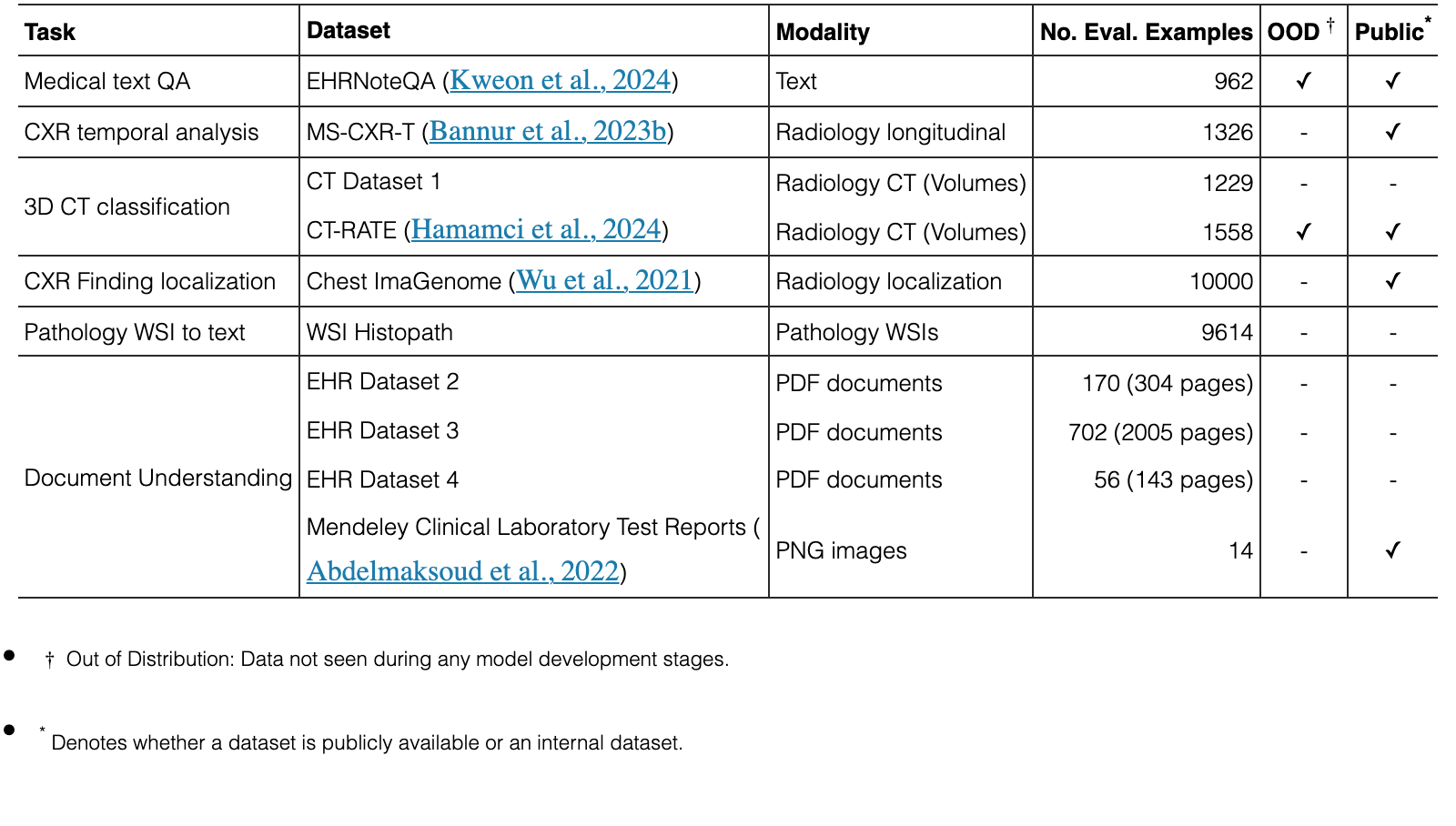
For training MedGemma 1.5, the vision encoder of MedGemma 1 (MedSigLIP) (Sellergren et al., 2025; Zhai et al., 2023) was frozen, and the language decoder underwent additional pretraining (supervised finetuning of the LLM) to build upon the initial baseline capabilities. We incorporated both the text and interleaved imaging data from the original Gemma mixture as well as the new medical domain image-text paired data as summarized in Table 1. These datasets were utilized via a combination of additional pretraining, distillation, and reinforcement learning as indicated in the table and described below.
We added new modalities for radiology and new data for dermatology and histopathology, and EHR/lab report understanding. Specifically, to train MedGemma 1.5 4B, the internal dermatology dataset was expanded to include additional examples from the open (CC-0) components of the ISIC 2017 and 2018 datasets (Gutman et al., 2016; Codella et al., 2018; Tschandl et al., 2018) as well as an internal set of images from a large hospital in Japan (adding to the dermatology datasets used previously and described in Yang et al. (2024)). Additionally, internal radiography datasets–CXR-IND1, CT Dataset 1, and MRI Dataset 1–were also added to pretraining.
The knowledge acquired from pretraining was refined in the post-training stage through a combination of distillation and reinforcement learning (RL), and we used the same recipes as Gemma 3, but with additional medical data for both steps. Distillation here means we sample 256 teacher model logits per token, weighted by teacher probabilities. The student learns the teacher’s distribution within these samples via cross-entropy loss (Team et al., 2025).
The distillation process for MedGemma 1.5 was augmented by incorporating additional domain-specific teacher models in addition to an improved large instruction-tuned (IT) teacher. For example, to enhance high-dimensional medical imaging capabilities, we trained supplementary teachers on CT Dataset 1, MRI-Dataset 1, and Internal histopathology. As indicated in Table 1, additional Distill and/or RL training was applied across multiple modalities–including radiology, dermatology, and whole slide pathology imaging–to further align the model’s performance with clinical visual tasks. To enabled improved document understanding, we distilled on EHRQA (synthetic records created by Synthea (Walonoski et al., 2018).) as well as EHR datasets 2, 3, 4, and 5.
Since the image encoder can only process 2D RGB images, 3D CT and MR image volumes were preprocessed to sequences of individual 2D axial images, each of which was rescaled to the image encoder’s input dimension of of pixels.
We capped the number of axial slices per query to a maximum of 85 during training and evaluation (which amount to 21,760 vision tokens), to stay below a total of 32K tokens when including the radiology report indication (included in the prompt) and the findings (target), in order to keep the memory requirements during training manageable. Slices could originate from multiple z-stacked volumes per study. Inclusion criteria for each of these volumes were: a) a maximum of 512\penalty\ \times 512 pixels per slice, b) axial orientation, c) slices with the same thickness, and d) at least five slices. For CT studies, these included volumes with different reconstruction kernels originating from the same scan, and for MRI studies volumes representing different sequences and contrasts, including T1-weighted (T1w), T2-weighted (T2w), GRE, and SWI. For computational efficiency, if the final z-stacked volume had more then 85 slices in total, we sampled slices equidistantly across the z-axis (of the stacked volume).
Regarding the mapping of voxel values, as with our previous work (Sellergren et al., 2025) for single slice CT images, we employed multi-channel windowing to map raw Hounsfield Units (HU) to RGB values, since the image encoder was trained only with 256 different intensity values per channel. Specifically, we used the following CT window mappings:
- •
Red Channel (-1024, 1024): Given the heterogeneity of our training data (spanning brain, chest, and abdomen), this wide window ensures that morphological boundaries, from air-filled lung parenchyma to dense cortical bone, remain visible across all anatomical regions.
- •
Green Channel (-135, 215): Since the Green channel contributes most significantly to luminance in standard image processing, we mapped the complex soft-tissue data here to leverage the encoder’s sensitivity to texture in visceral organs and mediastinal structures.
- •
Blue Channel (0, 80): This narrow, high-contrast window highlights subtle attenuations in brain parenchyma (gray/white matter differentiation) and acute intracranial hemorrhages, as well as vascular calcifications.
By contrast, since voxel values of MR images are all relative and no physiological windows exist, no windowing was applied, similar to the SigLip encoder (Sellergren et al., 2025). Instead we normalized them per volume using min-max normalization, and set the R, G, and B channels to the same value.
The histopathology WSI preprocessing pipeline was designed to convert WSIs into a sequence of representative tissue patches suitable for multi-modal learning. To ensure computational efficiency and relevance, we restricted patch extraction to tissue-containing regions. A tissue mask was generated for each slide at a low resolution (5x magnification). We employed a custom multi-stage tissue segmentation algorithm (Ahmed et al., 2025) operating in the HSV (Hue, Saturation, Value) color space.
For each WSI, a single optical magnification level was stochastically selected for patch extraction, with probabilities set to approximate uniformity across standard diagnostic levels: P(5x)=0.34, P(10x)=0.33, and P(20x)=0.33. The high-resolution tissue mask was downscaled to match the target extraction stride. Non-overlapping patches of size 896 by 896 pixels were extracted from a regular grid defined by the tissue mask, with the stride equal to the patch size. To maintain a fixed sequence length for downstream models, we enforced a cap of 126 patches per slide (leading to 32256 vision tokens) via random subsampling without replacement. Note that the number of images we used for long vision tasks differ for CT and WSI due to having different-length text inputs alongside them (i.e. CT had longer text inputs compared to WSI). Crucially, the original spatial ordering of the selected patches was preserved to retain relative positional context. The resulting patches were encoded as PNG images and stored alongside the slide caption. We chose a higher number of images per query than for CT and MRI examples above (126 versus 85), since fewer tokens were needed to encode image captions.
This processing is applied to an internal pathology dataset with a total of 335,825 whole slide images-text pairs for pretraining, distillation, and RL (on token-level ROUGE-L) as well as the 9,614 pairs used for evaluation.
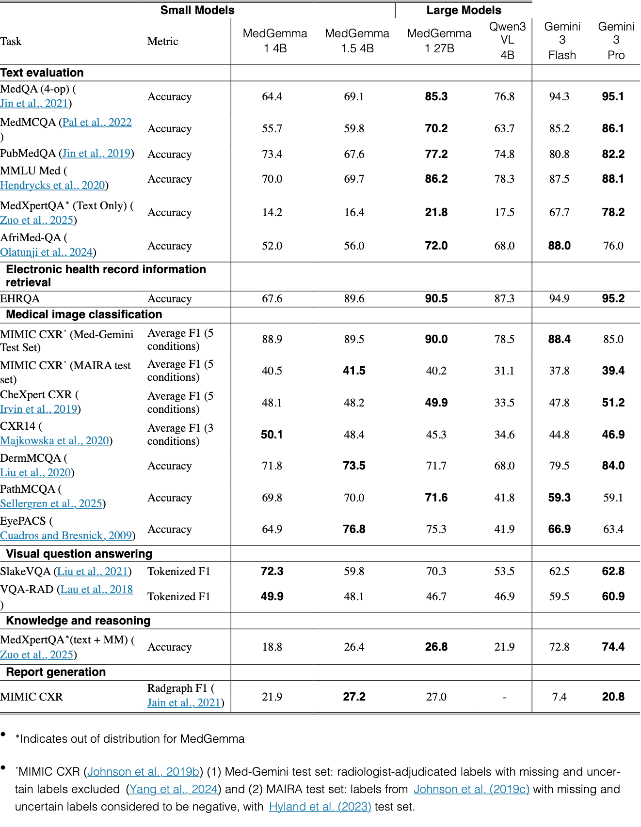
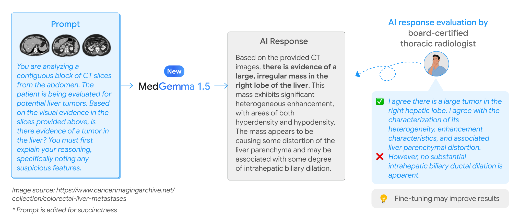
Results of evaluations for original tasks from Sellergren et al. (2025) are shown in Table 3 and results of evaluations on new tasks are shown in Table 4 and Figure 2.
| Small Models | Large Models | ||||||
| Task | Metric | MedGemma 1 4B | MedGemma 1.5 4B | MedGemma 1 27B | Qwen3 VL 4B | Gemini 3 Flash | Gemini 3 Pro |
| Text evaluation | |||||||
| MedQA (4-op) (Jin et al., 2021) | Accuracy | 64.4 | 69.1 | 85.3 | 76.8 | 94.3 | 95.1 |
| MedMCQA (Pal et al., 2022) | Accuracy | 55.7 | 59.8 | 70.2 | 63.7 | 85.2 | 86.1 |
| PubMedQA (Jin et al., 2019) | Accuracy | 73.4 | 67.6 | 77.2 | 74.8 | 80.8 | 82.2 |
| MMLU Med (Hendrycks et al., 2020) | Accuracy | 70.0 | 69.7 | 86.2 | 78.3 | 87.5 | 88.1 |
| MedXpertQA∗ (Text Only) (Zuo et al., 2025) | Accuracy | 14.2 | 16.4 | 21.8 | 17.5 | 67.7 | 78.2 |
| AfriMed-QA (Olatunji et al., 2024) | Accuracy | 52.0 | 56.0 | 72.0 | 68.0 | 88.0 | 76.0 |
| Electronic health record information retrieval | |||||||
| EHRQA | Accuracy | 67.6 | 89.6 | 90.5 | 87.3 | 94.9 | 95.2 |
| Medical image classification | |||||||
| MIMIC CXR⋄ (Med-Gemini Test Set) | Average F1 (5 conditions) | 88.9 | 89.5 | 90.0 | 78.5 | 88.4 | 85.0 |
| MIMIC CXR⋄ (MAIRA test set) | Average F1 (5 conditions) | 40.5 | 41.5 | 40.2 | 31.1 | 37.8 | 39.4 |
| CheXpert CXR (Irvin et al., 2019) | Average F1 (5 conditions) | 48.1 | 48.2 | 49.9 | 33.5 | 47.8 | 51.2 |
| CXR14 (Majkowska et al., 2020) | Average F1 (3 conditions) | 50.1 | 48.4 | 45.3 | 34.6 | 44.8 | 46.9 |
| DermMCQA (Liu et al., 2020) | Accuracy | 71.8 | 73.5 | 71.7 | 68.0 | 79.5 | 84.0 |
| PathMCQA (Sellergren et al., 2025) | Accuracy | 69.8 | 70.0 | 71.6 | 41.8 | 59.3 | 59.1 |
| EyePACS (Cuadros and Bresnick, 2009) | Accuracy | 64.9 | 76.8 | 75.3 | 41.9 | 66.9 | 63.4 |
| Visual question answering | |||||||
| SlakeVQA (Liu et al., 2021) | Tokenized F1 | 72.3 | 59.8 | 70.3 | 53.5 | 62.5 | 62.8 |
| VQA-RAD (Lau et al., 2018) | Tokenized F1 | 49.9 | 48.1 | 46.7 | 46.9 | 59.5 | 60.9 |
| Knowledge and reasoning | |||||||
| MedXpertQA∗(text + MM) (Zuo et al., 2025) | Accuracy | 18.8 | 26.4 | 26.8 | 21.9 | 72.8 | 74.4 |
| Report generation | |||||||
| MIMIC CXR | Radgraph F1 (Jain et al., 2021) | 21.9 | 27.2 | 27.0 | - | 7.4 | 20.8 |
View original table
| Small Models | Large Models | |||||||||
| Task | Metric | MedGemma 1 4B | MedGemma 1.5 4B | MedGemma 1 27B | Qwen3 VL 4B | Gemma 3 4B | Gemma 3 27B | Gemini 3.0 Flash | Gemini 3.0 Pro | External SOTA |
| Text Only | ||||||||||
| EHRNoteQA | Accuracy | 79.4 | 80.4 | 90.7 | 90.6 | 78.0 | 90.3 | 93.9 | 95.0 | 95.15-97.16(1) |
| Document Understanding | ||||||||||
| EHR Dataset 2 | Macro F1 | 78 | 91 | 76 | - | 84 | 93 | 92 | 93 | - |
| EHR Dataset 3 | Macro F1 | 50 | 71 | 66 | - | 61 | 74 | 74 | 90 | - |
| EHR Dataset 4 | Macro F1 | 25 | 64 | 5 | - | 41 | 52 | 82 | 81 | - |
| Mendeley Clinical Laboratory Test Reports | Macro F1 | 85 | 85 | 69 | - | 83 | 89 | 89 | 90 | - |
| CXR Analysis | ||||||||||
| Chest ImaGenome (Localization) | Mean IoU | 3.1 | 38.0 | 16.0 | 8.7 | 5.7 | 8.1 | 38.5 | 39.1 | 30.7-34.4 (2) |
| MS-CXR-T† (Temporal) | Macro-Accuracy | 61.1 | 65.7 | 50.1 | 53.5 | 59.0 | 52.7 | 67.3 | 62.9 | 68.5(3) |
| High-Dimension Image Analysis | ||||||||||
| CT Dataset 1 (3D CT) | Accuracy | 58.2 | 61.1 | 57.8 | 52.8 | 54.5 | 55.7 | 62.9 | 61.0 | - |
| MRI Dataset 1 (3D MRI) | Accuracy | 51.3 | 64.7 | 57.4 | 49.6 | 51.1 | 50.5 | 60.3 | 55.5 | - |
| WSI Histopath | ROUGE-L | 2.2 | 49.4 | 4.1 | ? | 2.3 | 3.2 | 13.9 | 12.2 | 49.8(4) |
View original table
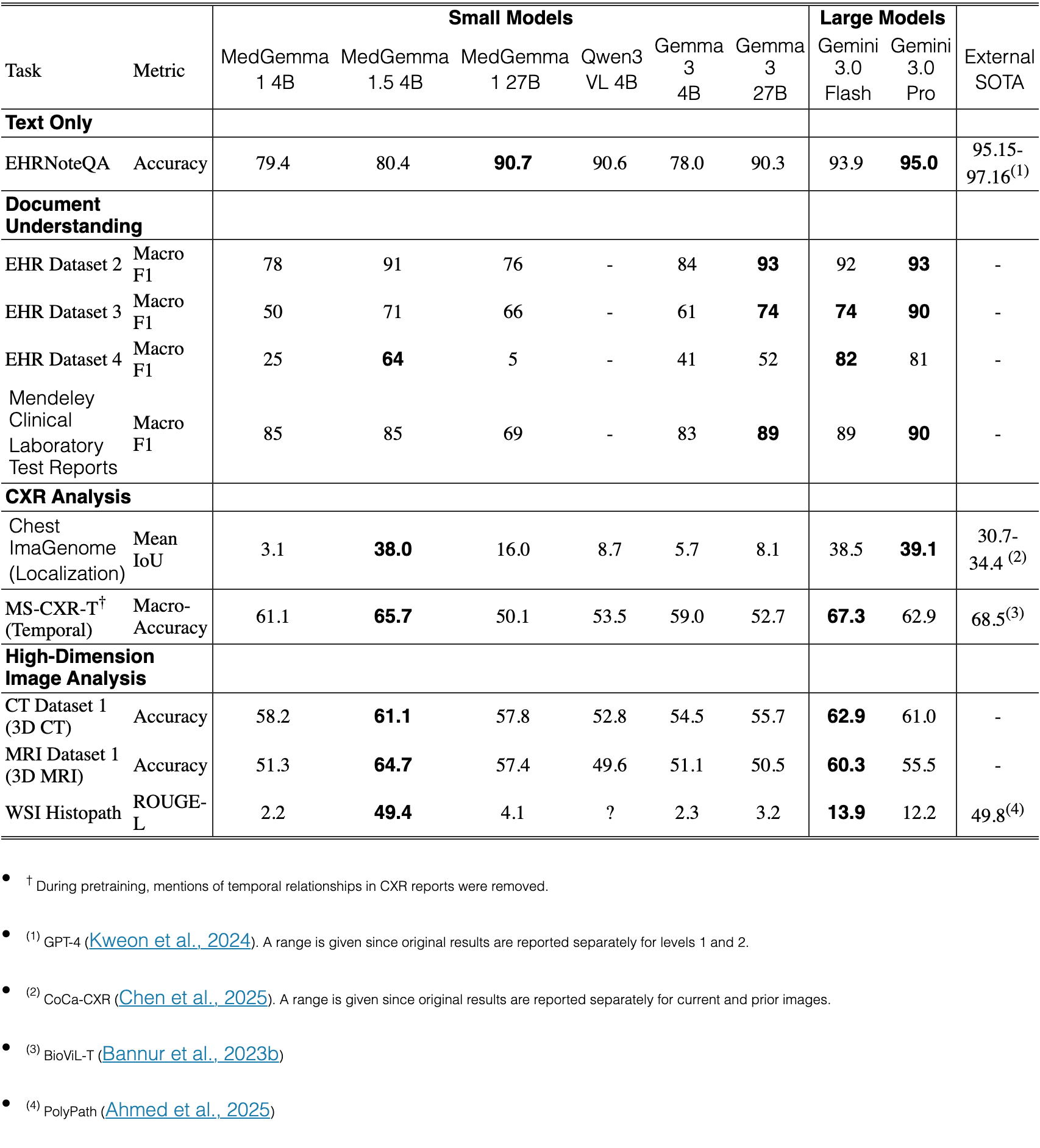
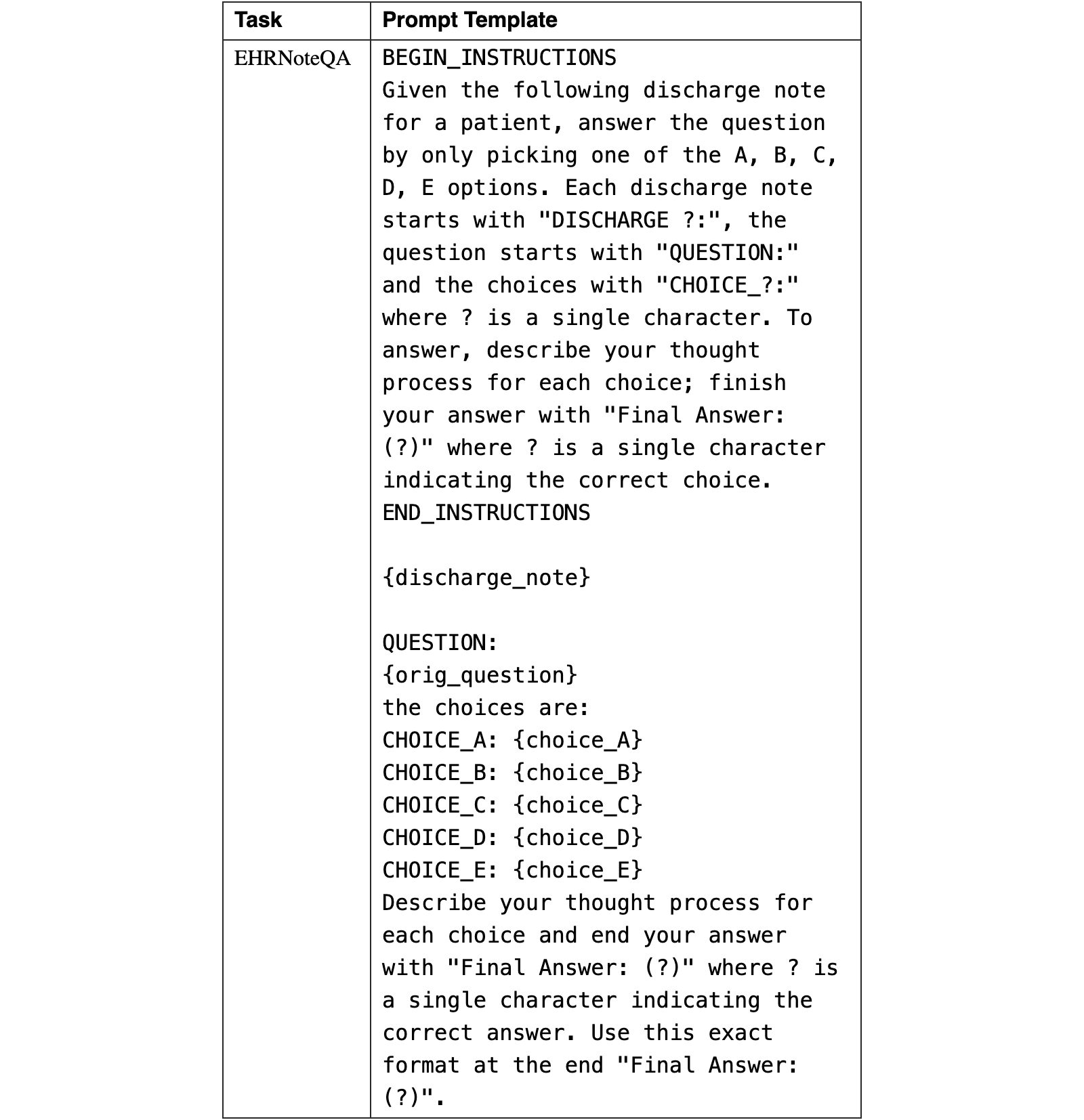
Unless reported otherwise, all evaluations that we performed consisted of a single inference run per example. For MedGemma 1.5 evaluations, a temperature of 0.0 was used on all evaluations based on informal measurements of tune set performance. For other baseline models, the default temperature was used. Temperature 0.0 was kept for new dataset evaluations of MedGemma 1 for consistency. For evaluations of all other models, on all datasets, each model’s default temperature and top-k were used. Note that results may vary with other choices of temperature and top-k. All evaluations were conducted using data sources or splits of data sources that were completely held out from MedGemma training. Prompts were updated for MedGemma 1.5 as summarized in Appendix B. Note that changes to prompts sometimes had a significant effect on benchmark performance and further optimization of prompts is likely possible.
Various prompts were updated and standardized compared to evaluations in Sellergren et al. (2025). In particular, for similar tasks, like MCQ, we now now utilize the same initial instruction prompt to be more consistent. Updated prompts are shown in Appendix B. Similar to the previous MedGemma models, we manually optimized prompts for MedGemma 1.5 on the training and validation splits to find prompts that worked well for evaluations. We briefly summarize the previous evaluation datasets here. See Sellergren et al. (2025) for further details.
For chest X-ray classification, performance was measured via F1-score across three datasets: MIMIC-CXR (Johnson et al., 2019b; Johnson et al., 2019a; Goldberger et al., 2000b), CheXpert (Irvin et al., 2019), and the ChestX-ray14 (CXR14) (Wang et al., 2017) dataset. On MIMIC-CXR, we evaluated five common lung conditions (atelectasis, cardiomegaly, consolidation, edema, and pleural effusion). Two test sets were used for MIMIC-CXR: a Med-Gemini Test Set (Yang et al., 2024), using radiologist-adjudicated labels instead of the original ones, and treating only explicitly 0-labeled conditions as negatives, i.e. leaving out non-mentioned or uncertain conditions, and a MAIRA test set using case selection from Hyland et al. (2023) and original labels from Johnson et al. (2019c) (treating uncertainty as negative). On ChestX-ray14, we evaluated three conditions–lung opacity, pneumothorax, and fracture–on radiologist-adjudicated labels from Majkowska et al. (2020). For CheXpert, we evaluated on all 14 labeled observations (Irvin et al., 2019) using the original labels.
For image-based MCQ, we evaluated accuracy on DermMCQA (Liu et al., 2020), PathMCQA (Sellergren et al., 2025; Jaroensri et al., 2022; Nagpal et al., 2019; Nagpal et al., 2020; Sadhwani et al., 2021), and EyePACS (Cuadros and Bresnick, 2009). DermMCQA (Liu et al., 2020) is an internal dermatology dataset consisting of one image per patient from 1996 patients. There are 136 different skin conditions in total, with ground truth diagnoses provided by dermatologists based on the images and metadata. We generated 4-option multiple choice questions (MCQs) using random distractors for each image. PathMCQA is an internal histopathology dataset, with a test split comprising of 450 patches from 354 whole slide images across breast, lung, prostate, lymph, and cervical specimens. Identification, grading, and sub-typing tasks were formulated as 4–9 option MCQs with ground truth from board-certified pathologists (Sellergren et al., 2025). On the EyePACS fundus image dataset (Cuadros and Bresnick, 2009), we evaluated against clinically determined, 5-class diabetic retinopathy severity grades, formatted as 5-option MCQs, for 3614 de-identified images, each originating from a different patient.
For general visual question answering of medical images, we evaluated average tokenized F1 on SLAKE (Liu et al., 2021) and VQA-RAD (Lau et al., 2018). SLAKE is a dataset of aggregated CT, MRI, and X-Ray images and questions of body parts (neck, pelvis, abdomen, head and chest). We used the default splits for SLAKE. VQA-RAD is a dataset of radiology images and questions of head, chest, and abdomen. For VQA-RAD, we used splits from Yang et al. (2024) instead of the original splits in order to prevent contamination of the test split.
We also reported a radiology report generation metric (RadGraph F1 (Jain et al., 2021)) on the 912-image MIMIC-CXR test set used in Tanno et al. (2024) and Yang et al. (2024).
For general medical text MCQ, we also evaluated accuracy on the publicly available test splits for MedQA, MedMCQA, PubMedQA, MMLU medical subcategories, AfriMed-QA, and MedXpertQA (Zuo et al., 2025). Note that MedXpertQA is out of distribution.
On CT and MR images we evaluated MedGemma and comparable models on the classification of common conditions.
The test split of the internal CT Dataset 1 consisted of head, chest, and abdominal/pelvis acquisitions, Models were evaluated on their ability to detect the following conditions: cardiac calcification, suspicious lung nodules (chest), aortic aneurysm, renal calculus, tumors, appendicitis (abdomen/pelvis), and hemorrhage (head). To extract binary ground truth labels (indicating presence or absence within the volume), we employed a multi-stage pipeline: (1) RegEx-based screening for positive mentions, (2) Gemini-based extraction, and (3) final manual review of a random subset by a US board-certified cardiothoracic radiologist. A condition was labeled ’absent’ if the report explicitly ruled it out or omitted any mention of it. The MRI Dataset 1 test split comprised brain, knee, and abdomen acquisitions. Labels were extracted using the same pipeline for the following conditions: acute infarct, hemorrhage, and multiple sclerosis (brain); meniscal tears and fractures (knee); and liver disease and pancreatic lesions (abdomen). Both internal datasets were sub-sampled without replacement to ensure a balanced distribution of positive and negative cases per condition. Image volumes were preprocessed as described in Section 2.3.1.
We also evaluated CT performance on the (internal) validation split of the public CT-RATE dataset (Hamamci et al., 2024), comprising of 1,564 non-contrast chest CT acquisitions from 1304 patients. As with the internal dataset, we measured macro accuracy across the binary prediction of 18 different conditions and abnormalities, respectively, while using the original labels provided with the dataset, which mostly cover common lung and cardiology conditions as described in (Hamamci et al., 2024). We preprocessed CT volumes in the same manner as described above, although not starting with raw volumes, but ones resampled to , as provided by (Hamamci et al., 2024).
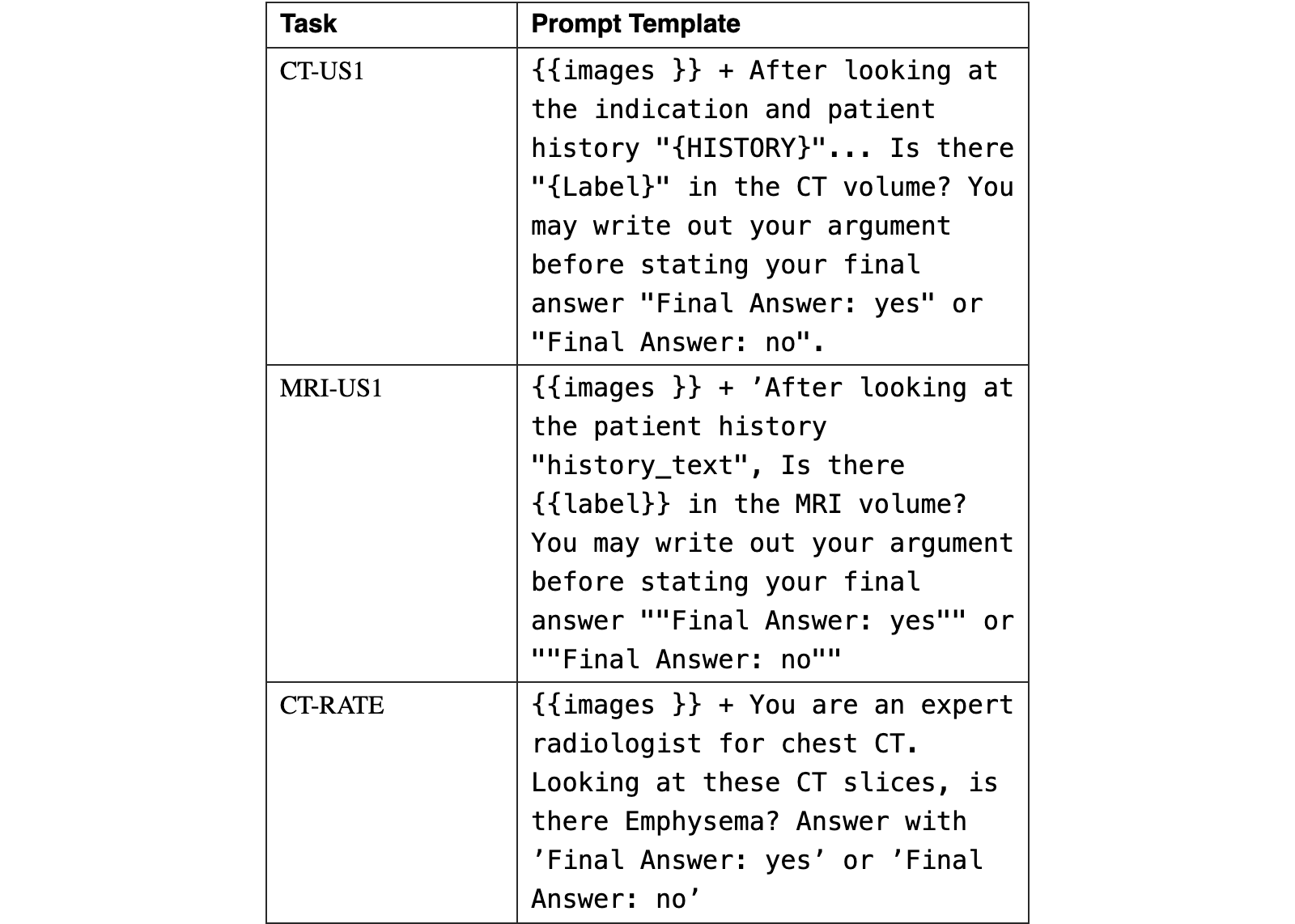
During evaluation, models were queried for each condition separately. Prompts consisted of the sequence of selected image slices interleaved with slice indices (e.g., SLICE {index}), followed by a binary question regarding the condition’s presence. For the internal datasets, the prompt also included the patient history from the original report (see Table 13). Because the models produced generative text rather than class probabilities, evaluation was based on binary presence/absence answers. We first computed performance metrics for each condition independently—using Accuracy for the balanced internal sets and F1 score for the imbalanced CT-RATE dataset—before calculating the Macro-Accuracy and Macro-F1 scores, respectively, to provide an aggregate measure of model performance. Note that CT-RATE results are included in Appendix A.
We evaluated using the ROUGE metric against the final diagnosis sections of the original pathology reports, having previously showed that this metric provides good correlation with pathologist scoring of image-text pairs for the same task (Ahmed et al., 2025). Each WSI was processed and preprocessed (with the patch extraction methods specified in Section 2.3.2) and provided as input along with the text specimen label (e.g. “colon biopsy”). For this analysis, only single WSI-text pairs are used.
To assesses the model’s capacity for temporal reasoning within longitudinal medical imaging, we evaluated on MS-CXR-T (Bannur et al., 2023a; Bannur et al., 2023b; Goldberger et al., 2000a). This task required the model to analyze pairs of chest X-rays—specifically a prior and a current study—to determine the trajectory of five specific cardiopulmonary pathologies: consolidation, edema, pleural effusion, pneumonia, and pneumothorax. The evaluation pipeline combined two radiographs with a structured text prompt, where for every image pair, the model is prompted to return one of three potential classes: (A) Improved, (B) Stable, or (C) Worsened. Performance is quantified using the macro accuracy metric to account for class imbalance (Bannur et al., 2023b).
To assess the model’s ability to localize anatomical regions of interest, we evaluated on Chest ImaGenome (Wu et al., 2021). We utilized a bounding box evaluation protocol. The model was prompted to generate coordinates of 2D bounding boxes for specific anatomical structures identified in the query. The primary metric for evaluating localization performance was the Intersection over Union (IoU). For a predicted bounding box and a ground truth bounding box , the IoU is defined as in Wu et al. (2021).
The model was provided with an input frontal chest X-ray image and a text prompt. The prompt contained specific instructions to output a JSON list of objects, where each object is defined by a label and a 2D bounding box coordinate set ‘[y0, x0, y1, x1]‘. The coordinates were requested to be normalized to the range , with representing the top-left corner and the bottom-right corner.
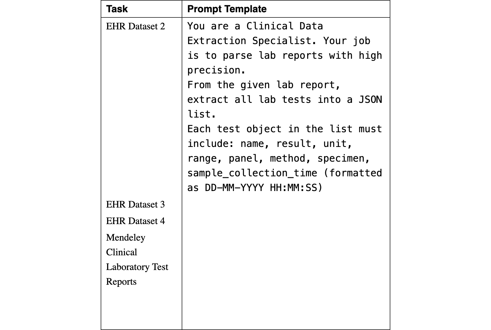
MedGemma 1.5 was tuned and evaluated on structured data extraction from multimodal laboratory reports, converting key attributes from document images and PDFs (rendered into an image via an open-source library) into a structured JSON format. This task may be considered a prerequisite for downstream standardization and interoperability tasks such as LOINC (Logical Observation Identifiers Names and Codes) mapping or FHIR (Fast Healthcare Interoperability Resources) resource generation.
To ensure robust generalization, we curated a composite dataset designed to reflect the complexities of real-world clinical data. The corpus inputs consist of medical laboratory reports provided as document images (e.g., PNG, JPEG) and Portable Document Formats (PDFs). The scope is specifically limited to pathology lab tests. The datasets encompass both digital-native reports and scanned documents, the latter introducing challenges such as noise, variable illumination, and rotational artifacts inherent to manual digitization.
The target JSON objects are structured to reflect the hierarchical and relational data of the source documents, focusing on the following data: name, result, unit, specimen, method, and sample collection time. F1 performance is calculated using a multi-phase label matcher algorithm to pair predicted labels with ground truth counterparts, followed by a metric calculation component that computes overall and granular (per-parameter) metrics: precision, recall, and F1 scores.
The required output for this task is a structured JSON object that accurately reflects the hierarchical and relational data of the source document, maintaining accurate key-value pairings for all extracted entities. This processing was applied to all EHR dataset 2, EHR dataset 3, EHR dataset 4, and Mendeley Clinical Laboratory Test Reports.
To assess the model’s capability in clinical reasoning over real-world electronic health records, we utilized the EHRNoteQA benchmark (Kweon et al., 2024). This dataset comprises 962 question-answer pairs derived from discharge summaries in the MIMIC-IV database, covering diverse clinical topics such as treatment plans, diagnostics, and patient history. Each instance involves analyzing accumulated discharge summaries for a specific patient to answer clinically relevant questions.
While the benchmark supports both open-ended and multiple-choice formats, our evaluation focused exclusively on the multiple-choice question (MCQ) setting. For each query, the model was presented with the patient’s discharge notes, a question, and five answer choices (A through E) and overall accuracy was assessed.
The release of MedGemma 1.5 marks an important step for open-source medical artificial intelligence. Its performance across diverse benchmarks demonstrates that a single, efficient 4B parameter model can not only retain competency across established text-based benchmarks but also generalize to complex tasks in high dimensional and longitudinal imaging. Notably, due to the improved distillation and RL, we see that the model is able to actually improve performance on multiple text-based benchmarks compared to 1.0, including 5% accuracy on MedQA and 8% on MedXperQA (multimodal). The model’s ability to process native modalities with spatial and temporal depth—synthesizing evidence across non-contiguous volumetric scans, whole-slide digital pathology, and longitudinal imaging—demonstrates that smaller LLMs can make progress towards understanding of 3D anatomy and disease progression.
Beyond strong benchmark performance, these architectural improvements establish MedGemma 1.5 as a highly practical foundation for developers. Innovations like the enhanced anatomical localization and multi-timepoint analysis provide out-of-the-box spatiotemporal awareness, while its robust EHR and Lab Report parsing capabilities enable medical-specific OCR use-cases. Importantly, these out-of-the-box functionalities are designed as foundational data processing tools, distinct from automated clinical decision-making or the practice of medicine. Rather than engineering complex multimodal pipelines from scratch, developers and researchers can leverage this efficient baseline as a versatile starting point to fine-tune bespoke clinical applications, accelerating the deployment of accessible, next-generation healthcare tools.
To contextualize MedGemma 1.5 within the broader ecosystem of open-weights models, we compared its performance against Qwen3 VL 4B, a similarly sized state-of-the-art multimodal model. The comparison reveals distinct design philosophies. Qwen3 VL 4B demonstrates superior performance on general text-based biomedical knowledge tasks, such as MedQA. However, MedGemma 1.5 significantly outperforms Qwen3 VL 4B in specialized clinical vision tasks that require domain-specific visual capabilities. On all vision tasks, MedGemma 1.5 achieved higher performance. This divergence highlights the utility of MedGemma’s specialized post-training and distillation pipeline; while generalist models excel at knowledge retrieval, the MedGemma lineage retains an edge in the interpretation of nuanced multimodal reasoning (e.g. via its superiority in multimodal MedXpertQA and all Medical image classification tasks).
In our comparative analysis of novel evaluation tasks, we aimed to provide a broad perspective by including external baselines such as Qwen3 VL 4B. Some evaluations were not possible to perform with Qwen3 due to logistical constraints within our current internal evaluation framework. Future work may involve extending this framework to accommodate the distinct inference protocols of the Qwen architecture.
Compared to MedGemma 1, we observed trade-offs inherent to the expansion of model capabilities. As MedGemma 1.5 became more of a “medical generalist”, we noted minor regressions in specific legacy benchmarks, such as SLAKE (Liu et al., 2021) and VQA-RAD (Lau et al., 2018). However, we note that these benchmarks have their own limitations in terms of quality as evaluations rely on token overlap, and the ground truth answers are not standardized. We believe this new model is more generally useful at medical imaging due to the improved performance on high-dimensional imaging and bounding box localization. Developers seeking maximum performance on narrower tasks can bridge these gaps through targeted fine-tuning.
MedGemma 1.5 significantly expands the utility of the original model by advancing beyond standard 2D tasks to tackle high-dimensional, spatiotemporal modalities such as 3D radiology, pathology whole-slide imaging, and longitudinal imaging sequences. Despite these complex new capabilities and added document-understanding features, the model retains the computational and cost efficiency of a 4B parameter architecture. By releasing this highly capable, multimodally-aware foundation as an open resource, we aim to empower the developer community with an even more useful starting point to bridge the gap between academic benchmarks and impactful, real-world clinical applications.
The models have been released openly at the main Google Health AI Developer Foundations site at https://goo.gle/hai-def. Further details specifically about the MedGemma collection of models can be found at https://goo.gle/medgemma.
Technical Leads
Andrew Sellergren
Fereshteh Mahvar
Core contributors
Chufan Gao
Timo Kohlberger
Fayaz Jamil
Madeleine Traverse
Alberto Tono
Bashir Sadjad
Lin Yang
Charles Lau
Liron Yatziv
Tiffany Chen
Bram Sterling
Kenneth Philbrick
Richa Tiwari
Yun Liu
Madhuram Jajoo
Chandrashekar Sankarapu
Swapnil Vispute
Harshad Purandare
Abhishek Bijay Mishra
Contributors
Sam Schmidgall
Tao Tu
Anil Palepu
Chunjong Park
Tim Strother
Rahul Thapa
Yong Cheng
Preeti Singh
Kat Black
Sponsors
Yossi Matias
Katherine Chou
Avinatan Hassidim
Kavi Goel
Joelle Barral
Tris Warkentin
Leads
Shravya Shetty
Dale Webster
Sunny Virmani
David F. Steiner
Can Kirmizibayrak
Daniel Golden
Last author
Co-first author
Many teams from both Google Research and Google DeepMind collaborated extensively on this project. We thank Ellery Wulczyn and Greg Corrado for their feedback and insight, which significantly enhanced this report.
Sections of this manuscript was drafted using Gemini 2.5 Pro and Gemini 3 Pro and further refined via human editors over multiple rounds. Final manual checks were performed to ensure content accuracy. The authors take full responsibility for the content.
References
- Abdelmaksoud et al. (2022) E. Abdelmaksoud, A. Gadallah, and A. Asad Clinical Laboratory Test Reports. Mendeley Data. External Links: Document, Link Cited by: Table 2.SourceSource
- Ahmed et al. (2025) F. Ahmed, L. Yang, T. Jaroensri, A. Sellergren, Y. Matias, A. Hassidim, G. S. Corrado, D. R. Webster, S. Shetty, S. Prabhakara, et al. Polypath: adapting a large multimodal model for multi-slide pathology report generation. arXiv preprint arXiv:2502.10536. Cited by: §2.3.2, 5th item, §3.2.2.
- Bannur et al. (2023a) S. Bannur, S. Hyland, Q. Liu, F. Pérez-García, M. Ilse, D. Coelho de Castro, B. Boecking, H. Sharma, K. Bouzid, A. Schwaighofer, M. T. Wetscherek, H. Richardson, T. Naumann, J. Alvarez Valle, and O. Oktay MS-CXR-T: Learning to Exploit Temporal Structure for Biomedical Vision-Language Processing. PhysioNet. Note: Version 1.0.0 External Links: Document, Link Cited by: §3.2.3.SourceSource
- Bannur et al. (2023b) S. Bannur, S. Hyland, Q. Liu, F. Perez-Garcia, M. Ilse, D. C. Castro, B. Boecking, H. Sharma, K. Bouzid, A. Thieme, et al. Learning to exploit temporal structure for biomedical vision-language processing. In Proceedings of the IEEE/CVF Conference on Computer Vision and Pattern Recognition, pp. 15016–15027. Cited by: Table 2, 4th item, §3.2.3.
- Chen et al. (2025) Y. Chen, S. Xu, A. Sellergren, Y. Matias, A. Hassidim, S. Shetty, D. Golden, A. L. Yuille, and L. Yang CoCa-cxr: co ntrastive ca ptioners learn strong temporal structures for chest x-ray vision-language understanding. In International Conference on Medical Image Computing and Computer-Assisted Intervention, pp. 78–88. Cited by: 3rd item.
- Codella et al. (2018) N. C. F. Codella, D. Gutman, M. E. Celebi, B. Helba, M. A. Marchetti, S. W. Dusza, A. Kalloo, K. Liopyris, N. Mishra, H. Kittler, and A. Halpern Skin lesion analysis toward melanoma detection: a challenge at the 2017 international symposium on biomedical imaging (isbi), hosted by the international skin imaging collaboration (isic). External Links: 1710.05006, Link Cited by: §2.1.Source
- Cuadros and Bresnick (2009) J. Cuadros and G. Bresnick EyePACS: an adaptable telemedicine system for diabetic retinopathy screening. Journal of diabetes science and technology 3 (3), pp. 509–516. Cited by: §3.1, Table 3.
- Goldberger et al. (2000a) A. Goldberger, L. Amaral, L. Glass, J. Hausdorff, P. C. Ivanov, R. Mark, and H. E. Stanley PhysioBank, physiotoolkit, and physionet: components of a new research resource for complex physiologic signals. Circulation 101 (23), pp. e215–e220. Note: Online; RRID:SCR_007345 Cited by: §3.2.3.
- Goldberger et al. (2000b) A. L. Goldberger, L. A. Amaral, L. Glass, J. M. Hausdorff, P. C. Ivanov, R. G. Mark, J. E. Mietus, G. B. Moody, C. Peng, and H. E. Stanley PhysioBank, physiotoolkit, and physionet: components of a new research resource for complex physiologic signals. circulation 101 (23), pp. e215–e220. Cited by: §3.1.
- Gutman et al. (2016) D. Gutman, N. C. Codella, E. Celebi, B. Helba, M. Marchetti, N. Mishra, and A. Halpern Skin lesion analysis toward melanoma detection: a challenge at the international symposium on biomedical imaging (isbi) 2016, hosted by the international skin imaging collaboration (isic). arXiv preprint arXiv:1605.01397. Cited by: §2.1.
- Hamamci et al. (2024) I. E. Hamamci, S. Er, C. Wang, F. Almas, A. G. Simsek, S. N. Esirgun, I. Dogan, O. F. Durugol, B. Hou, S. Shit, et al. Developing generalist foundation models from a multimodal dataset for 3d computed tomography. arXiv preprint arXiv:2403.17834. Cited by: Appendix A, Table 2, §3.2.1.
- Hendrycks et al. (2020) D. Hendrycks, C. Burns, S. Basart, A. Zou, M. Mazeika, D. Song, and J. Steinhardt Measuring massive multitask language understanding. arXiv preprint arXiv:2009.03300. Cited by: Table 3.
- Hyland et al. (2023) S. L. Hyland, S. Bannur, K. Bouzid, D. C. Castro, M. Ranjit, A. Schwaighofer, F. Pérez-García, V. Salvatelli, S. Srivastav, A. Thieme, et al. MAIRA-1: a specialised large multimodal model for radiology report generation. arXiv preprint arXiv:2311.13668. Cited by: 2nd item, §3.1.
- Irvin et al. (2019) J. Irvin, P. Rajpurkar, M. Ko, Y. Yu, S. Ciurea-Ilcus, C. Chute, H. Marklund, B. Haghgoo, R. Ball, K. Shpanskaya, et al. Chexpert: a large chest radiograph dataset with uncertainty labels and expert comparison. In Proceedings of the AAAI conference on artificial intelligence, Vol. 33, pp. 590–597. Cited by: §3.1, Table 3.
- Jain et al. (2021) S. Jain, A. Agrawal, A. Saporta, S. Q. Truong, D. N. Duong, T. Bui, P. Chambon, Y. Zhang, M. P. Lungren, A. Y. Ng, et al. Radgraph: extracting clinical entities and relations from radiology reports. arXiv preprint arXiv:2106.14463. Cited by: §3.1, Table 3.
- Jaroensri et al. (2022) R. Jaroensri, E. Wulczyn, N. Hegde, T. Brown, I. Flament-Auvigne, F. Tan, Y. Cai, K. Nagpal, E. A. Rakha, D. J. Dabbs, et al. Deep learning models for histologic grading of breast cancer and association with disease prognosis. NPJ breast cancer 8 (1), pp. 113. Cited by: §3.1.
- Jin et al. (2021) D. Jin, E. Pan, N. Oufattole, W. Weng, H. Fang, and P. Szolovits What disease does this patient have? a large-scale open domain question answering dataset from medical exams. Applied Sciences 11 (14), pp. 6421. Cited by: Table 3.
- Jin et al. (2019) Q. Jin, B. Dhingra, Z. Liu, W. W. Cohen, and X. Lu Pubmedqa: a dataset for biomedical research question answering. arXiv preprint arXiv:1909.06146. Cited by: Table 3.
- Johnson et al. (2019a) A. Johnson, T. Pollard, R. Mark, S. Berkowitz, and S. Horng MIMIC-CXR database (version 2.0. 0). PhysioNet. Cited by: §3.1.
- Johnson et al. (2019b) A. E. Johnson, T. J. Pollard, S. J. Berkowitz, N. R. Greenbaum, M. P. Lungren, C. Deng, R. G. Mark, and S. Horng MIMIC-cxr, a de-identified publicly available database of chest radiographs with free-text reports. Scientific data 6 (1), pp. 317. Cited by: 2nd item, §3.1.
- Johnson et al. (2019c) A. Johnson, M. Lungren, Y. Peng, Z. Lu, R. Mark, S. Berkowitz, and S. Horng MIMIC-cxr-jpg - chest radiographs with structured labels. PhysioNet. External Links: Document, Link Cited by: 2nd item, §3.1.SourceSource
- Kweon et al. (2024) S. Kweon, J. Kim, H. Kwak, D. Cha, H. Yoon, K. H. Kim, J. Yang, S. Won, and E. Choi EHRNoteQA: an llm benchmark for real-world clinical practice using discharge summaries. Note: PhysioNet, version 1.0.1 External Links: Link Cited by: Table 2, 2nd item, §3.3.Source
- Lau et al. (2018) J. J. Lau, S. Gayen, A. Ben Abacha, and D. Demner-Fushman A dataset of clinically generated visual questions and answers about radiology images. Scientific data 5 (1), pp. 1–10. Cited by: §3.1, Table 3, §4.
- Liu et al. (2021) B. Liu, L. Zhan, L. Xu, L. Ma, Y. Yang, and X. Wu SLAKE: a semantically-labeled knowledge-enhanced dataset for medical visual question answering. In 2021 IEEE 18th International Symposium on Biomedical Imaging (ISBI), pp. 1650–1654. Cited by: §3.1, Table 3, §4.
- Liu et al. (2020) Y. Liu, A. Jain, C. Eng, D. H. Way, K. Lee, P. Bui, K. Kanada, G. de Oliveira Marinho, J. Gallegos, S. Gabriele, et al. A deep learning system for differential diagnosis of skin diseases. Nature medicine 26 (6), pp. 900–908. Cited by: §3.1, Table 3.
- Majkowska et al. (2020) A. Majkowska, S. Mittal, D. F. Steiner, J. J. Reicher, S. M. McKinney, G. E. Duggan, K. Eswaran, P. Cameron Chen, Y. Liu, S. R. Kalidindi, et al. Chest radiograph interpretation with deep learning models: assessment with radiologist-adjudicated reference standards and population-adjusted evaluation. Radiology 294 (2), pp. 421–431. Cited by: §3.1, Table 3.
- Nagpal et al. (2019) K. Nagpal, D. Foote, Y. Liu, P. C. Chen, E. Wulczyn, F. Tan, N. Olson, J. L. Smith, A. Mohtashamian, J. H. Wren, et al. Development and validation of a deep learning algorithm for improving gleason scoring of prostate cancer. NPJ digital medicine 2 (1), pp. 48. Cited by: §3.1.
- Nagpal et al. (2020) K. Nagpal, D. Foote, F. Tan, Y. Liu, P. C. Chen, D. F. Steiner, N. Manoj, N. Olson, J. L. Smith, A. Mohtashamian, et al. Development and validation of a deep learning algorithm for gleason grading of prostate cancer from biopsy specimens. JAMA oncology 6 (9), pp. 1372–1380. Cited by: §3.1.
- Olatunji et al. (2024) T. Olatunji, C. Nimo, A. Owodunni, T. Abdullahi, E. Ayodele, M. Sanni, C. Aka, F. Omofoye, F. Yuehgoh, T. Faniran, et al. AfriMed-qa: a pan-african, multi-specialty, medical question-answering benchmark dataset. arXiv preprint arXiv:2411.15640. Cited by: Table 3.
- Pal et al. (2022) A. Pal, L. K. Umapathi, and M. Sankarasubbu Medmcqa: a large-scale multi-subject multi-choice dataset for medical domain question answering. In Conference on health, inference, and learning, pp. 248–260. Cited by: Table 3.
- Sadhwani et al. (2021) A. Sadhwani, H. Chang, A. Behrooz, T. Brown, I. Auvigne-Flament, H. Patel, R. Findlater, V. Velez, F. Tan, K. Tekiela, et al. Comparative analysis of machine learning approaches to classify tumor mutation burden in lung adenocarcinoma using histopathology images. Scientific reports 11 (1), pp. 16605. Cited by: §3.1.
- Sellergren et al. (2025) A. Sellergren, S. Kazemzadeh, T. Jaroensri, A. Kiraly, M. Traverse, T. Kohlberger, S. Xu, F. Jamil, C. Hughes, C. Lau, et al. Medgemma technical report. arXiv preprint arXiv:2507.05201. Cited by: Appendix B, §1, §2.1, §2.3.1, §2.3.1, §2, §3.1, §3.1, Table 3, §3.
- Tanno et al. (2024) R. Tanno, D. Barrett, A. Sellergren, S. Ghaisas, S. Dathathri, A. See, J. Welbl, K. Singhal, S. Azizi, T. Tu, et al. Consensus, dissensus and synergy between clinicians and specialist foundation models in radiology report generation. arXiv preprint arXiv:2311.18260. Cited by: §3.1.
- Team et al. (2025) G. Team, A. Kamath, J. Ferret, S. Pathak, N. Vieillard, R. Merhej, S. Perrin, T. Matejovicova, A. Ramé, M. Rivière, L. Rouillard, T. Mesnard, G. Cideron, J. Grill, S. Ramos, E. Yvinec, M. Casbon, E. Pot, I. Penchev, G. Liu, F. Visin, K. Kenealy, L. Beyer, X. Zhai, A. Tsitsulin, R. Busa-Fekete, A. Feng, N. Sachdeva, B. Coleman, Y. Gao, B. Mustafa, I. Barr, E. Parisotto, D. Tian, M. Eyal, C. Cherry, J. Peter, D. Sinopalnikov, S. Bhupatiraju, R. Agarwal, M. Kazemi, D. Malkin, R. Kumar, D. Vilar, I. Brusilovsky, J. Luo, A. Steiner, A. Friesen, A. Sharma, A. Sharma, A. M. Gilady, A. Goedeckemeyer, A. Saade, A. Feng, A. Kolesnikov, A. Bendebury, A. Abdagic, A. Vadi, A. György, A. S. Pinto, A. Das, A. Bapna, A. Miech, A. Yang, A. Paterson, A. Shenoy, A. Chakrabarti, B. Piot, B. Wu, B. Shahriari, B. Petrini, C. Chen, C. L. Lan, C. A. Choquette-Choo, C. Carey, C. Brick, D. Deutsch, D. Eisenbud, D. Cattle, D. Cheng, D. Paparas, D. S. Sreepathihalli, D. Reid, D. Tran, D. Zelle, E. Noland, E. Huizenga, E. Kharitonov, F. Liu, G. Amirkhanyan, G. Cameron, H. Hashemi, H. Klimczak-Plucińska, H. Singh, H. Mehta, H. T. Lehri, H. Hazimeh, I. Ballantyne, I. Szpektor, I. Nardini, J. Pouget-Abadie, J. Chan, J. Stanton, J. Wieting, J. Lai, J. Orbay, J. Fernandez, J. Newlan, J. Ji, J. Singh, K. Black, K. Yu, K. Hui, K. Vodrahalli, K. Greff, L. Qiu, M. Valentine, M. Coelho, M. Ritter, M. Hoffman, M. Watson, M. Chaturvedi, M. Moynihan, M. Ma, N. Babar, N. Noy, N. Byrd, N. Roy, N. Momchev, N. Chauhan, N. Sachdeva, O. Bunyan, P. Botarda, P. Caron, P. K. Rubenstein, P. Culliton, P. Schmid, P. G. Sessa, P. Xu, P. Stanczyk, P. Tafti, R. Shivanna, R. Wu, R. Pan, R. Rokni, R. Willoughby, R. Vallu, R. Mullins, S. Jerome, S. Smoot, S. Girgin, S. Iqbal, S. Reddy, S. Sheth, S. Põder, S. Bhatnagar, S. R. Panyam, S. Eiger, S. Zhang, T. Liu, T. Yacovone, T. Liechty, U. Kalra, U. Evci, V. Misra, V. Roseberry, V. Feinberg, V. Kolesnikov, W. Han, W. Kwon, X. Chen, Y. Chow, Y. Zhu, Z. Wei, Z. Egyed, V. Cotruta, M. Giang, P. Kirk, A. Rao, K. Black, N. Babar, J. Lo, E. Moreira, L. G. Martins, O. Sanseviero, L. Gonzalez, Z. Gleicher, T. Warkentin, V. Mirrokni, E. Senter, E. Collins, J. Barral, Z. Ghahramani, R. Hadsell, Y. Matias, D. Sculley, S. Petrov, N. Fiedel, N. Shazeer, O. Vinyals, J. Dean, D. Hassabis, K. Kavukcuoglu, C. Farabet, E. Buchatskaya, J. Alayrac, R. Anil, Dmitry, Lepikhin, S. Borgeaud, O. Bachem, A. Joulin, A. Andreev, C. Hardin, R. Dadashi, and L. Hussenot Gemma 3 technical report. External Links: 2503.19786, Link Cited by: §2.2, §2.Source
- Tschandl et al. (2018) P. Tschandl, C. Rosendahl, and H. Kittler The ham10000 dataset, a large collection of multi-source dermatoscopic images of common pigmented skin lesions. Scientific Data 5 (1). External Links: ISSN 2052-4463, Link, Document Cited by: §2.1.SourceSource
- Walonoski et al. (2018) J. Walonoski, M. Kramer, J. Nichols, A. Quina, C. Moesel, D. Hall, C. Duffett, K. Dube, T. Gallagher, and S. McLachlan Synthea: an approach, method, and software mechanism for generating synthetic patients and the synthetic electronic health care record. Journal of the American Medical Informatics Association 25 (3), pp. 230–238. External Links: Document Cited by: §2.2.Source
- Wang et al. (2017) X. Wang, Y. Peng, L. Lu, Z. Lu, M. Bagheri, and R. M. Summers Chestx-ray8: hospital-scale chest x-ray database and benchmarks on weakly-supervised classification and localization of common thorax diseases. In Proceedings of the IEEE conference on computer vision and pattern recognition, pp. 2097–2106. Cited by: §3.1.
- Wu et al. (2021) J. Wu, N. Agu, I. Lourentzou, A. Sharma, J. Paguio, J. S. Yao, E. C. Dee, W. Mitchell, S. Kashyap, A. Giovannini, et al. Chest imagenome dataset. Physio Net. Cited by: Table 1, Table 2, §3.2.4.
- Yang et al. (2024) L. Yang, S. Xu, A. Sellergren, T. Kohlberger, Y. Zhou, I. Ktena, A. Kiraly, F. Ahmed, F. Hormozdiari, T. Jaroensri, et al. Advancing multimodal medical capabilities of gemini. arXiv preprint arXiv:2405.03162. Cited by: §1, §2.1, 2nd item, §3.1, §3.1, §3.1.
- Zhai et al. (2023) X. Zhai, B. Mustafa, A. Kolesnikov, and L. Beyer Sigmoid loss for language image pre-training. In Proceedings of the IEEE/CVF international conference on computer vision, pp. 11975–11986. Cited by: §2.1.
- Zuo et al. (2025) Y. Zuo, S. Qu, Y. Li, Z. Chen, X. Zhu, E. Hua, K. Zhang, N. Ding, and B. Zhou Medxpertqa: benchmarking expert-level medical reasoning and understanding. arXiv preprint arXiv:2501.18362. Cited by: §3.1, Table 3, Table 3.
We additionally evaluated a portion of our models on the CT-RATE dataset (Hamamci et al., 2024), where we process accordingly (without resampling) per Section 2.3.1 with results summarized in Table 5. Unlike specialized, custom-built CT architectures that are optimized to yield multi-label predictions in a single forward pass, applying generalist vision-language models to this high-dimensional task required a more granular inference strategy. Specifically, our framework necessitated querying the model 18 times per condition to accurately parse the diagnostic signal. This iterative interrogation—while essential for leveraging the generative capabilities of the model—introduced a substantial computational bottleneck compared to the efficient, one-shot inference typical of dedicated CT classifiers. Consequently, due to the lengthy runtime associated with this eighteen-fold increase in query volume, we limited this evaluation to a representative subset of our models. We chose to evaluate on the most relevant models, as well as Gemini 3.0 Flash (which has shown to be competitive with Gemini 3.0 Pro in multimodal tasks).
The results presented in Table 5 highlight a significant divergence in performance between domain-specialized and generalist architectures on high-dimensional medical imaging tasks. Most notably, the MedGemma 4B models (versions 1 and 1.5) demonstrates superior zero-shot generalization capabilities compared to the general-purpose Gemini 3.0 Flash model. Despite the CT-RATE dataset being out-of-distribution (OOD) for the MedGemma training curriculum, these models achieved much higher Macro F1 scores. This disparity likely stems from the domain-specific pretraining of MedGemma, enabling a robust "medical prior," enabling more effective feature extraction from volumetric data even when the specific dataset distribution is novel.
| Task | Metric | MedGemma 1 4B | MedGemma 1.5 4B | Gemini 3.0 Flash |
| CT-RATE∗ (3D CT) | Macro F1 | 23.5 | 26.9 | 8.5 |
View original table

| Type | Benchmark | MedGemma 1 4B | MedGemma 1.5 4B | Gemma 3 4B§ | MedGemma 1 27B | Gemma 3 27B§ |
| Text-only | MMLU Pro | 39.1 | 33.8 | 43.6 | 60.2 | 67.5 |
View original table
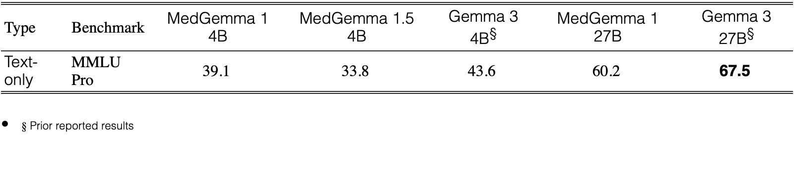
Table 6 shows a degradation in general knowledge reasoning compared to both its predecessor, MedGemma 1 4B and the foundational Gemma 3 4B. This decline suggests a clear tradeoff in the 4B parameter class, indicating that the intensive fine-tuning required to specialize the model for imaging may have resulted a diminished capacity for out-of-domain, general-purpose tasks. Still, we believe that this trade off is worth it for the large performance gains in the multimodal medical domain.
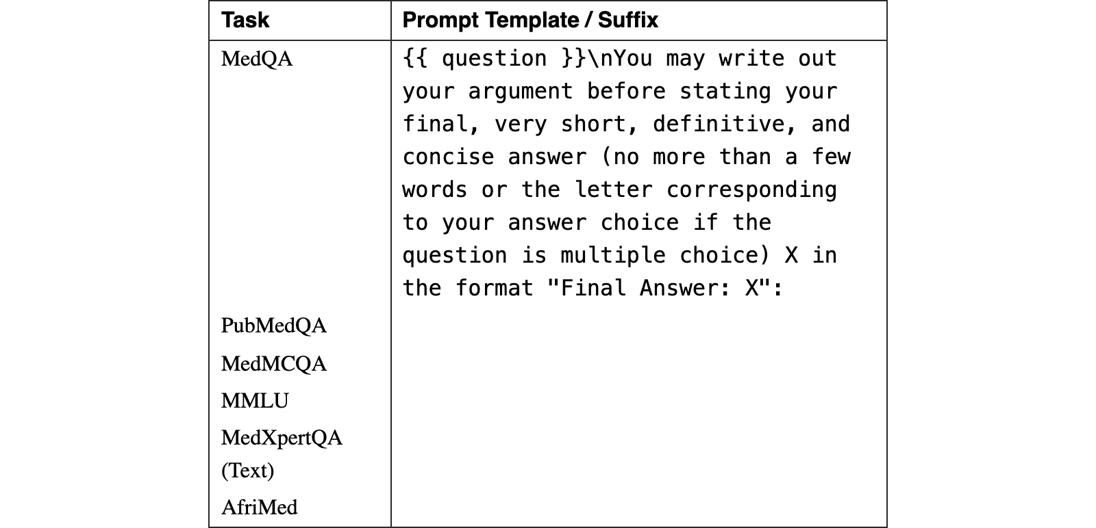
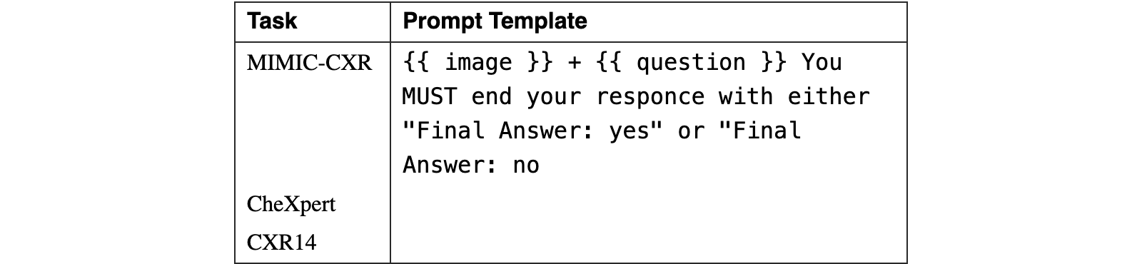
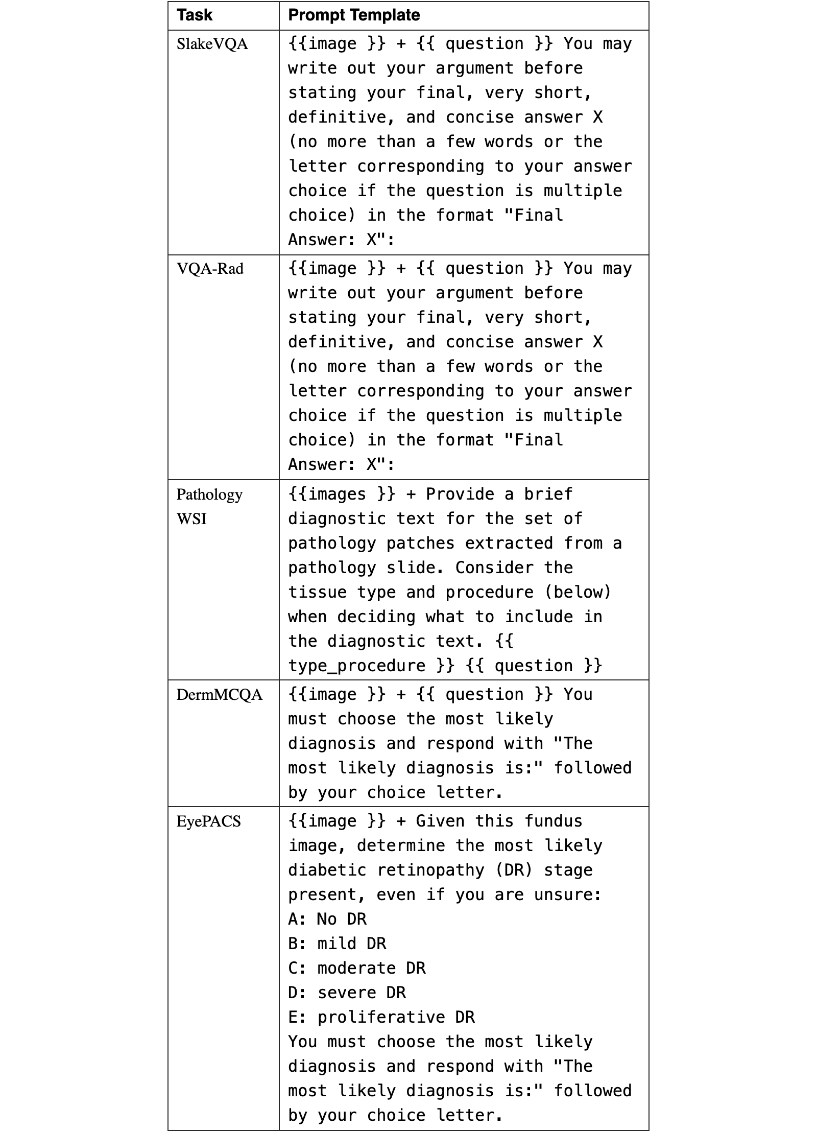
This section contains a complete list of all prompts for MedGemma 1.5. Prompts not listed here, such as prompts for running EHRQA, are identical to those used in Sellergren et al. (2025).
Please note that MedGemma models do not have a system instruction (system prompt). For general models, we apply the following system instruction to all evaluations that require radiology or chest X-ray interpretation: MS CXRT, SlakeVQA, VQA-Rad, Chest ImaGenome (Localization) "You are a helpful radiology assistant.". For all other benchmarks, we apply the following "You are a helpful medical assistant.".
For MedGemma 1.5, we also turn thinking on for certain benchmarks to encourage reasoning. Specificaly, we turn thinking on by appending "SYSTEM INSTRUCTION: think silently if needed." to the system prompt for the following benchmarks: MedQA, MedMCQA, EHRNoteQA, PubMedQA, MMLU Med, MedXpertQA (Text Only), and AfriMed-QA.
| Task | Prompt Template / Suffix |
| MedQA | {{ question }}\nYou may write out your argument before stating your final, very short, definitive, and concise answer (no more than a few words or the letter corresponding to your answer choice if the question is multiple choice) X in the format "Final Answer: X": |
| PubMedQA | |
| MedMCQA | |
| MMLU | |
| MedXpertQA (Text) | |
| AfriMed |
View original table
| Task | Prompt Template |
| MIMIC-CXR | {{ image }} + {{ question }} You MUST end your responce with either "Final Answer: yes" or "Final Answer: no |
| CheXpert | |
| CXR14 |
View original table
| Task | Prompt Template |
| SlakeVQA | {{image }} + {{ question }} You may write out your argument before stating your final, very short, definitive, and concise answer X (no more than a few words or the letter corresponding to your answer choice if the question is multiple choice) in the format "Final Answer: X": |
| VQA-Rad | {{image }} + {{ question }} You may write out your argument before stating your final, very short, definitive, and concise answer X (no more than a few words or the letter corresponding to your answer choice if the question is multiple choice) in the format "Final Answer: X": |
| Pathology WSI | {{images }} + Provide a brief diagnostic text for the set of pathology patches extracted from a pathology slide. Consider the tissue type and procedure (below) when deciding what to include in the diagnostic text. {{ type_procedure }} {{ question }} |
| DermMCQA | {{image }} + {{ question }} You must choose the most likely diagnosis and respond with "The most likely diagnosis is:" followed by your choice letter. |
| EyePACS | {{image }} + Given this fundus image, determine the most likely diabetic retinopathy (DR) stage present, even if you are unsure: A: No DR B: mild DR C: moderate DR D: severe DR E: proliferative DR You must choose the most likely diagnosis and respond with "The most likely diagnosis is:" followed by your choice letter. |
View original table
| Task | Prompt Template |
| EHRNoteQA | BEGIN_INSTRUCTIONS Given the following discharge note for a patient, answer the question by only picking one of the A, B, C, D, E options. Each discharge note starts with "DISCHARGE ?:", the question starts with "QUESTION:" and the choices with "CHOICE_?:" where ? is a single character. To answer, describe your thought process for each choice; finish your answer with "Final Answer: (?)" where ? is a single character indicating the correct choice. END_INSTRUCTIONS {discharge_note} QUESTION: {orig_question} the choices are: CHOICE_A: {choice_A} CHOICE_B: {choice_B} CHOICE_C: {choice_C} CHOICE_D: {choice_D} CHOICE_E: {choice_E} Describe your thought process for each choice and end your answer with "Final Answer: (?)" where ? is a single character indicating the correct answer. Use this exact format at the end "Final Answer: (?)". |
View original table
| Task | Prompt Template |
| EHR Dataset 2 | You are a Clinical Data Extraction Specialist. Your job is to parse lab reports with high precision. From the given lab report, extract all lab tests into a JSON list. Each test object in the list must include: name, result, unit, range, panel, method, specimen, sample_collection_time (formatted as DD-MM-YYYY HH:MM:SS) |
| EHR Dataset 3 | |
| EHR Dataset 4 | |
| Mendeley Clinical Laboratory Test Reports | |
View original table
| Task | Prompt Template |
| Chest ImaGenome (Localization) | {{image }} + Where is the {object}? |
View original table

| Task | Prompt Template |
| CT-US1 | {{images }} + After looking at the indication and patient history "{HISTORY}"... Is there "{Label}" in the CT volume? You may write out your argument before stating your final answer "Final Answer: yes" or "Final Answer: no". |
| MRI-US1 | {{images }} + ’After looking at the patient history "history_text", Is there {{label}} in the MRI volume? You may write out your argument before stating your final answer ""Final Answer: yes"" or ""Final Answer: no"" |
| CT-RATE | {{images }} + You are an expert radiologist for chest CT. Looking at these CT slices, is there Emphysema? Answer with ’Final Answer: yes’ or ’Final Answer: no’ |
View original table